Step 7. Quality control of cleaned bundles
Every participant and hemisphere is inspected after cleaning. The script converts each cleaned bundle to a tract-density image (TDI) with tckmap, selects the axial and coronal slices of greatest density, overlays the image on the mean b = 0 image, and writes one image per participant together with automatic flags (Table 1). Flags appear in the image title and are collected in qc_flags.csv.
Table 1
Automatic Flags
| Flag | Criterion |
|---|---|
LOW_COVERAGE | fewer than 50 TDI voxels |
HIGH_COVERAGE | more than 5,000 TDI voxels |
LOW_DENSITY | maximum density below 5 streamlines per voxel |
Script 07_visual_qc.py (118 lines)
#!/usr/bin/env python3
"""Step 7. Tract-density overlay of each cleaned bundle on the b0, with gross-failure flags.

Inputs:  $OUT/<subj>/tckgen/<TRACT>/<TRACT>_<CUTOFF>_cleaned.tck  (Step 6)
         $PROJECT/dwi/<subj>/nodif_brain_mask.nii.gz and mean_b0.nii.gz (optional)
Outputs: $OUT/qc/<TRACT>/<subj>_<TRACT>_qc.png and $OUT/qc/<TRACT>/qc_flags.csv
Run:     source 00_config.sh && python 07_visual_qc.py   (tckmap must be on PATH)
"""
import os
import subprocess
import sys
from pathlib import Path

# Agg backend, so the script also runs on a cluster node or over ssh with no display.
import matplotlib
matplotlib.use("Agg")
import matplotlib.pyplot as plt
import nibabel as nib
import numpy as np
import pandas as pd


# Read one variable exported by 00_config.sh, or stop with a message saying what to do.
def env(name):
    if name not in os.environ:
        sys.exit(f"{name} is not set: run `source 00_config.sh` first")
    return os.environ[name]


# CUTOFF stays a string because it only builds the file name Steps 5 and 6 used.
PROJECT = Path(env("PROJECT"))
OUT = Path(env("OUT"))
TRACT = env("TRACT")
CUTOFF = env("CUTOFF")
FORCE = os.environ.get("FORCE", "0") == "1"
SUBJECTS = Path(env("SUBJECTS_FILE")).read_text().split()

# Flag thresholds (Table 1 on the Step 7 page). They catch gross failures only and depend
# on voxel size and DILATE_VOX, so revisit them if your corridor differs from the example.
MIN_VOXELS = 50         # fewer occupied voxels: LOW_COVERAGE
MAX_VOXELS = 5000       # more occupied voxels: HIGH_COVERAGE (bundle leaves the pathway)
MIN_PEAK_DENSITY = 5    # peak streamlines per voxel below this: LOW_DENSITY

QC = OUT / "qc" / TRACT
QC.mkdir(parents=True, exist_ok=True)


# Turn the two numbers measured from the TDI into one flag; first hit wins, "" means pass.
def flag_for(n_voxels, peak):
    if n_voxels < MIN_VOXELS:
        return "LOW_COVERAGE"
    if n_voxels > MAX_VOXELS:
        return "HIGH_COVERAGE"
    if peak < MIN_PEAK_DENSITY:
        return "LOW_DENSITY"
    return ""


# Draw one panel, the b0 slice in grey with the density slice on top. np.rot90 puts
# anterior (axial) or superior (coronal) at the top; left-right is storage order, not L/R.
def overlay(ax, background, density, title):
    ax.imshow(np.rot90(background), cmap="gray")
    ax.imshow(np.rot90(np.ma.masked_where(density <= 0, density)), cmap="hot", alpha=0.7)
    ax.set_title(title)
    ax.axis("off")


# Every participant gets a row, so qc_flags.csv always lists the whole sample.
rows = []
for s in SUBJECTS:
    # The brain mask doubles as the tckmap template so the TDI lands on the diffusion grid.
    tck = OUT / s / "tckgen" / TRACT / f"{TRACT}_{CUTOFF}_cleaned.tck"
    template = PROJECT / "dwi" / s / "nodif_brain_mask.nii.gz"
    b0 = PROJECT / "dwi" / s / "mean_b0.nii.gz"
    png = QC / f"{s}_{TRACT}_qc.png"

    if not tck.exists():
        rows.append({"Subject": s, "Voxels": 0, "Peak_density": 0, "Flag": "MISSING"})
        print(f"[{s}] MISSING cleaned bundle")
        continue

    # tckmap's default output is the tract-density image (streamlines per voxel); the file
    # is scratch and is deleted once the array is in memory.
    tdi = QC / f"_{s}_tdi.nii.gz"
    subprocess.run(["tckmap", str(tck), str(tdi), "-template", str(template),
                    "-force", "-quiet"], check=True)
    density = nib.load(str(tdi)).get_fdata()
    tdi.unlink()

    n_voxels = int((density > 0).sum())
    peak = float(density.max())
    flag = flag_for(n_voxels, peak)
    rows.append({"Subject": s, "Voxels": n_voxels, "Peak_density": peak, "Flag": flag})
    print(f"[{s}] {n_voxels} voxels, peak {peak:.0f}  {flag}")

    # Flags were recomputed above; the picture is only redrawn when missing or FORCE=1.
    if png.exists() and not FORCE:
        continue
    background = nib.load(str(b0 if b0.exists() else template)).get_fdata()
    # Busiest axial and coronal slices; assumes x left-right, y front-back, z bottom-top.
    k_axial = int(np.argmax(density.sum(axis=(0, 1))))
    k_coronal = int(np.argmax(density.sum(axis=(0, 2))))
    fig, axes = plt.subplots(1, 2, figsize=(10, 5))
    overlay(axes[0], background[:, :, k_axial], density[:, :, k_axial], "Axial")
    overlay(axes[1], background[:, k_coronal, :], density[:, k_coronal, :], "Coronal")
    fig.suptitle(f"{s}  {TRACT}  cleaned" + (f"  [{flag}]" if flag else ""), fontsize=11)
    fig.tight_layout()
    fig.savefig(png, dpi=100, bbox_inches="tight")
    plt.close(fig)

# Anything with a non-empty Flag counts as flagged, so MISSING participants show up too.
flags = pd.DataFrame(rows)
flags.to_csv(QC / "qc_flags.csv", index=False)
flagged = flags[flags["Flag"] != ""]
print(f"\n{len(flagged)} of {len(flags)} participants flagged")
if len(flagged):
    print(flagged.to_string(index=False))
print(f"flags -> {QC / 'qc_flags.csv'}")
Criteria
A correct reconstruction shows the expected shape for the tract (for ventral tegmental area [VTA] → hippocampus, an arc from the ventral midbrain laterally into the medial temporal lobe), consistent shape across participants with variation in size, and no isolated voxel clusters, no density in the contralateral hemisphere or frontal lobe, and no empty tracts (Figures 1 to 3).
Figure 1
Cleaned Left Posterior VTA → Hippocampus Tract, One Participant
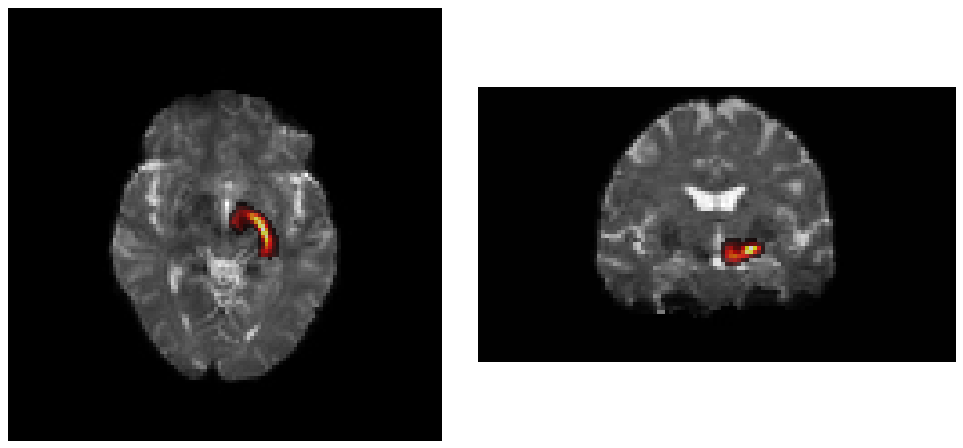
Note. Streamline density over the mean b = 0 image. Left: axial view. Right: coronal view. The same layout applies to Figures 2 and 3.
Figure 2
Cleaned Tract at the Low End of Retention
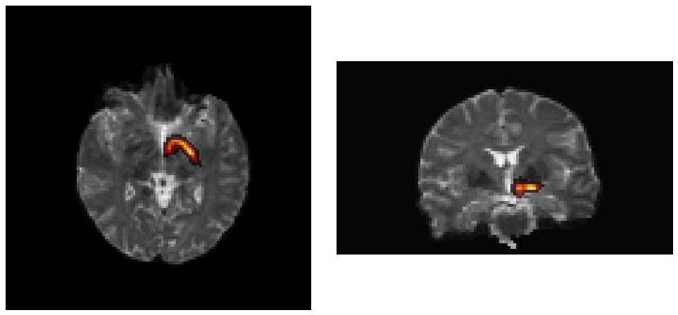
Note. About a quarter of the streamlines survived cleaning; the bundle is still well formed.
Figure 3
Cleaned Anterior VTA → Hippocampus Tract
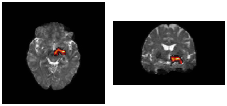
Note. Same participant as Figure 1.
Interactive review
The overlay images support a batch review. Flagged participants, and a sample of unflagged participants, should additionally be examined in a viewer with slice navigation, magnification and opacity control.
fsleyes "$PROJECT/dwi/$s/mean_b0.nii.gz" \
  "$OUT/$s/tckgen/$TRACT/${TRACT}_${CUTOFF}_cleaned.tck" -cm hot -a 70 &

mrview "$PROJECT/dwi/$s/mean_b0.nii.gz" \
  -tractography.load "$OUT/$s/tckgen/$TRACT/${TRACT}_${CUTOFF}_cleaned.tck" &
Comparison with the atlas
For a quantitative check, each participant's cleaned TDI can be warped to Montreal Neurological Institute (MNI) space with the inverse warp from Step 1 and compared with the atlas by Dice overlap against the 50% map, or by the fraction of the participant tract lying within the 25% probability map. This overlap should not be reported as validation of the atlas, because the reconstruction was constrained by it.
tckmap "$tck" "$tdi" -template "$PROJECT/dwi/$s/nodif_brain_mask.nii.gz" -force
fslmaths "$tdi" -thr 1 -bin "${tdi%.nii.gz}_bin.nii.gz"